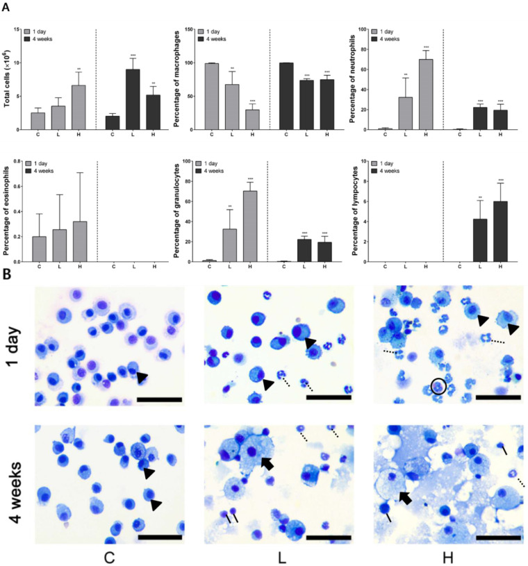
Figure 2.
Cytological analysis of bronchoalveolar lavage fluid (BALF) after intratracheal instillation of nickel oxide nanoparticles (NiO NPs). The number of total cells, percentage of macrophages, neutrophils, eosinophils, granulocytes, and lymphocytes is indicated (A). Diff–Quik staining images of immune cells in BALF after intratracheal instillation of NiO NPs. Macrophages (arrowhead) were observed after 1 day in C, L, and H. However, foamy macrophages (arrow) were observed only at 4 weeks in the L and H (B). Although neutrophils (dotted line) were recruited in the BALF of L and H, lymphocytes (solid line) were recruited at 4 weeks after instillation in BALF of L and H. Eosinophils (circle) were then observed 1 day after instillation in H from the cell image. Data are presented as mean ± standard deviation (SD) of each group. One-way analysis of variance (ANOVA) was performed for the comparison between the NiO NPs-treated and control groups with statistical significance indicated by ** p < 0.01 and *** p < 0.001. C: control group; L: low-dose group (50 cm2/rat); H: high-dose group (150 cm2/rat).

