Figure 2.
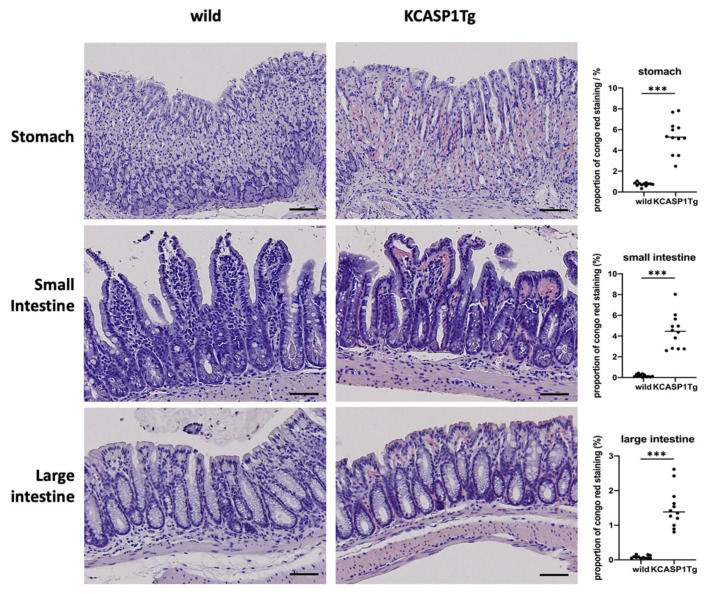
Histological analysis showed the amyloid deposition in KCASP1Tg mice. At 16 weeks of age, the stomach, small intestine, and large intestine were collected from the WT and KCASP1Tg mice and stained with Congo red. Compared with the WT mice, the KCASP1Tg mice had more severe amyloid deposition in the gastric mucosa and intestinal epithelial cells. Scale bar = 50 μm. There was a significant increase in the Congo red stain-positive area in the KCASP1Tg mice. All data were expressed as means ± SDs. *** p < 0.001 between KCASP1Tg and wild-type mice by Mann–Whitney test.

