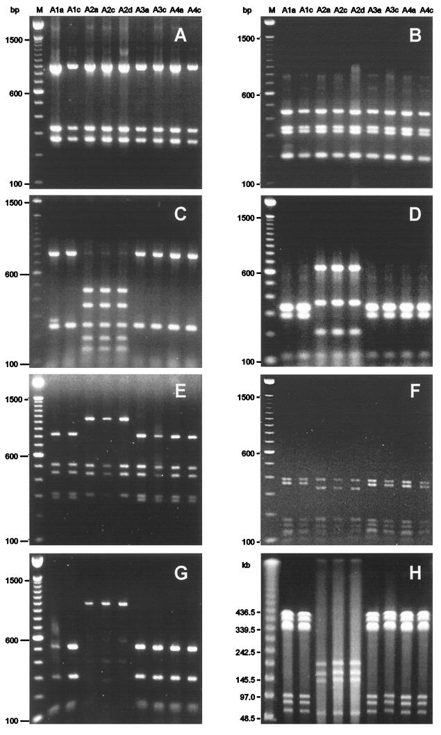
FIG. 1.
Representative molecular typing results for H. pylori isolates from family A. PCR-RFLP analyses of 16S rDNA fragments digested with HaeIII (A), 16S rDNA fragments digested with HinfI (B), flaA fragments digested with HhaI (C), flaA fragments digested with Sau3AI (D), ureAB fragments digested with HaeIII (E), lspA-glmA fragments digested with HhaI (F), and vacA fragments digested with HphI (G) and PFGE of genomic DNA digested with NotI (H) are shown. Lanes: M, size markers; A1a to A4c, H. pylori isolates, designated according to the origins of the isolates (A, family A; 1 to 4, family members; a, c, and d, antrum, corpus, and duodenum, respectively).

