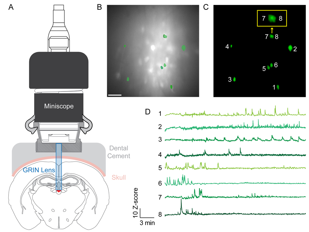
Figure 1.
Functional imaging of neuronal activity from a deep-brain region in vivo using a single-photon miniscope.
A) Schematic cross-sectional representation of a 500-μm GRIN lens inserted above the paraventricular thalamus (PVT) and miniscope attachment for imaging in freely moving mice. An adeno-associated virus with jGCaMP7f (AAV9-syn-FLEX-jGCaMP7f-WPRE; Addgene:104492-AAV9) was injected into the PVT of Drd2-Cre transgenic mice (MGI:3836635). B) Representative image depicting the maximum intensity per pixel from a motion-corrected video. Scale bar = 100 μm. C) Selected sample ROIs from (B) extracted by CNMF-E. (Inset outlined in yellow is a zoomed-in view of neurons 7 and 8, demonstrating the ability of CNMF-E to demix overlapping signals). D) Calcium traces extracted by CNMF-E corresponding to ROIs in (C). Permission to publish miniscope drawing granted by Doric Lenses Inc.

