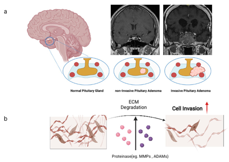
Figure 1.
(a) Pituitary gland located in the sella turcica shown in coronal sections as normal pituitary gland, benign pituitary adenoma, and as invasive pituitary adenoma. Internal carotid arteries adjacent to the pituitary are depicted as red circles. Cavernous sinuses are depicted in light blue. Note that aggressive PAs tend to circumvent the arteries. (b) Cellular mode of invasion into the brain tissue depends on degradation of extracellular matrix (ECM) molecules by proteinases of the metzincin family, namely MMPs and ADAMs. All images produced with Biorender (https://biorender.com (accessed on 6 November 2021)).

