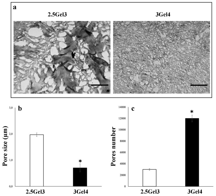
Figure 7.
(a) Optical microscope representative images of the 2.5Gel3 and 3Gel4 hydrogels for the pores’ morphological analysis. Image area = 25 µm2, scale bar 1 µm; (b) average pore size (µm) of the 2.5Gel3 and 3Gel4 hydrogels and (c) average pores number, as analyzed by ImageJ software; * p < 0.01.

