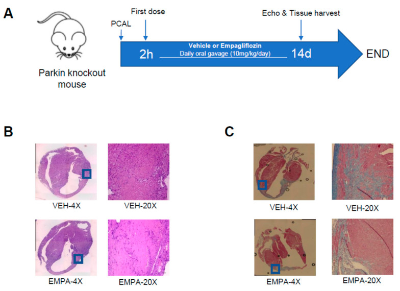
Figure 3.
EMPA limits infarct-mediated development of adverse remodeling in Parkin knockout mice. Age-matched WT mice underwent PCAL and 2 h later were given vehicle (0.5% w/v hydroxyethyl cellulose) or EMPA (10 mg/kg/day) via oral gavage daily. (A) Schematic of the protocol; (B) representative 4× and 20× magnification of heart sections stained with H&E of infarct border zone from 3 days after PCAL to show infiltration of immune cells; (C) representative 4× and 20× magnification of heart sections with Masson Trichrome staining 14 days after PCAL showing fibrosis.

