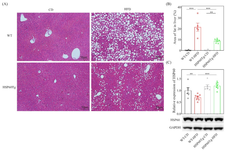
Figure 2.
A gain of HSP60 reduces hepatocellular steatosis in the context of chronic HFD for 26 weeks. Paraformaldehyde-fixed paraffin-embedded liver tissue was used to determine the abundance of lipid droplets with hematoxylin–eosin (HE) stain. (A) Representative HE stains image of each group. (B) Lipid droplet area quantified using ImageJ. (C) The expression of HSP60 in tissues from the HFD group was significantly lower than in tissues from the WT mice. Furthermore, overexpression of HSP60 was significantly greater in HSP60Tg-HFD mice compared with the WT-HFD group. Data collected from six fields of view of each specimen and five to seven specimens for each group are expressed as the mean ± SE. ** p < 0.01 and *** p < 0.001 between the indicated groups. WT, wild-type mice; HFD, high-fat diet; HSP60Tg, mice harboring overexpression of heat shock protein 60; CD, chow diet.

