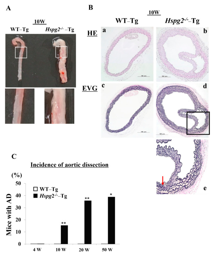
Figure 1.
Hspg2−/−-Tg mice frequently had an aortic dissection (AD). (A) Representative image of thoracic AD in Hspg2−/−-Tg mice at 10 weeks of age, with a possibility that it may occur to whole or part of the thoracic aortic tissue. (B) Representative image of hematoxylin/eosin (HE) staining (a,b) and Elastica van Gieson (EVG) staining (c–e) indicating the tear of elastic lamina of the medial wall (red arrow) (Scale bar = 100 µm). (C) Hspg2−/−-Tg mice had AD with a frequency of about 15.4% at 10 weeks of age (6/39), 35.8% at 20 weeks of age (5/16), and 38.9% at 50 weeks of age (7/18). (Fisher’s exact test, * p < 0.05, ** p < 0.01 vs. WT-Tg). Histological and morphological analyses of aortic tissue in Hspg2−/−-Tg mice at 1 weeks and 4 weeks of age are shown in supplementary Figure S1.

