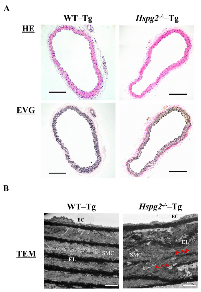
Figure 3.
Histological and morphological analyses of aortic tissue in Hspg2−/−-Tg mice indicated the absence of aortic dissection (AD). (A) HE staining and EVG staining showed no significant differences in the aortic tissue morphology of Hspg2−/−-Tg mice compared to that of WT-Tg, with no AD at 10 weeks of age (Scale bar = 100 µm, n = 6). (B) TEM analysis showed that the elastic lamina (EL) in Hspg2−/−-Tg mice was partially torn and thinner (red arrows) than that in the WT-Tg mice. (Scale bar = 5 µm, n = 3). These images are typical.

