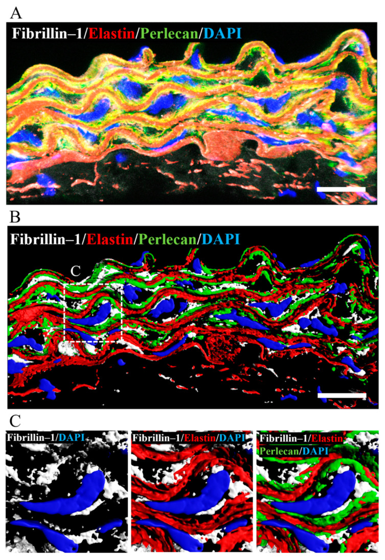
Figure 7.
Perlecan colocalized with fibrillin-1 and elastin in WT-Tg mice aorta. (A) Immunostaining was performed with anti-perlecan (green), -elastin (red), and -fibrillin-1 (white) antibodies. (B) The image is displayed in 3D using the IMARIS software after surface reconstruction of each labeling. Perlecan was localized along the elastic lamina. (C) Close up of the insert in B. Perlecan was localized at the intersection between elastin and fibrillin-1. (Scale bar = 20µm). The images for Hspg2−/−-Tg mice aorta are shown in supplementary Figure S2.

