Figure 3.
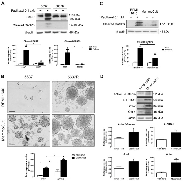
Paclitaxel sensitivity is reduced in 5637 cells grown as tumorspheres. (A) Western blots of PARP and cleaved caspase-3 in parental 5637 and paclitaxel-resistant 5637R cells treated with DMSO (vehicle) or 0.1 µM paclitaxel for 48 h. (B) Representative images of tumorspheres from parental 5637 and paclitaxel-resistant 5637R cells grown for 5 days in non-adherent conditions with RPMI 1640 or MammoCultTM. Bar, 100 µM. Histograms show the number of tumorspheres per well. (C) Western blot of cleaved caspase-3 in 5637 cells cultured in RPMI 1640 and adherent conditions or MammoCultTM and non-adherent conditions and treated with DMSO (vehicle) or 0.1 µM paclitaxel for 48 h. (D) Western blots of active β-catenin, ALDH1A1, Sox-2 and Oct-4 in 5637 cells cultured in RPMI 1640 and adherent conditions or MammoCultTM and non-adherent conditions. β-actin was used as a loading control. Histograms show the densitometric analysis of the indicated proteins. Data are presented as mean ± SD. * p value < 0.05 from Student’s t test (n ≥ 3). ns, not significant.

