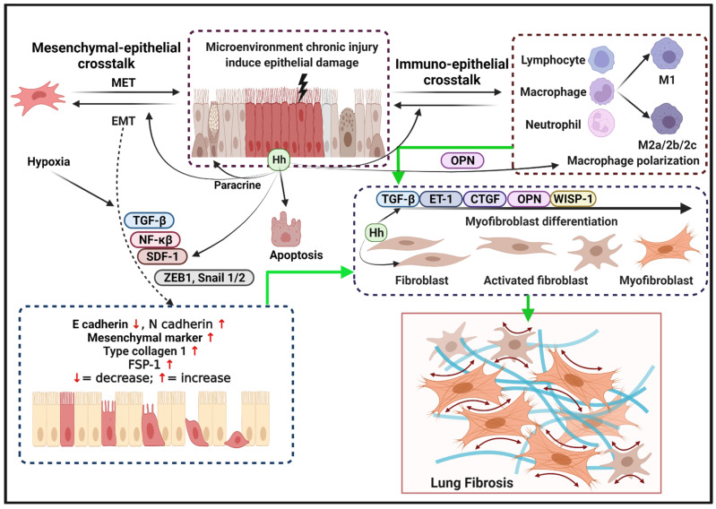
Figure 2.
Model of uncontrolled reactivation of Hh signaling in the pathogenesis of IPF. Microenvironment factors provoke repetitive epithelial injury followed by secretion of the Hh signaling pathway-regulated various products, such as TGF-β, SDF-1, Snail 1/2, OPN, ZEB-1, and NF-κβ. Epithelial cell Hh also warns neighboring cells via paracrine signals, induces apoptosis, and initiates crosstalk with immune and mesenchymal cells. Growth factor and microenvironment-induced epithelial and mesenchymal crosstalk promote the formation of EMT characterized by increased N-cadherin, mesenchymal markers, FSP1, and type I collagen. Hh signaling as one of activating transcriptional factors AECIIs regulates the immune response to ameliorate lung injury by undergoing EMT mechanism, promoting macrophage M2-associated inflammatory components, and fibroblasts recruitment to the injured site. Furthermore, Hh signaling pathway regulates myofibroblast differentiation and ECM production in parallel with other pro-fibrogenic proteins and cytokines, including CTGF, TGF-β, α-SMA, ET-1, OPN, and WISP-1. In conjunction with other pro-fibrogenic factors and cytokines, the Hh pathway regulates the accumulation of ECM-associated myofibroblast, collagen synthesis, and lung architecture is replaced with scar tissue fibrosis.

