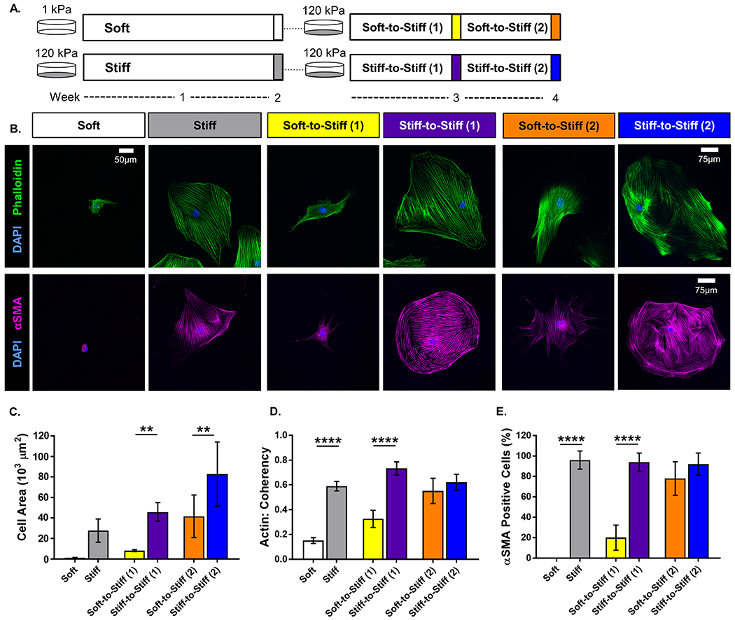
Figure 1.

Schematic of the in vitro experimental method timeline. Adipose-derived stem cells (ASCs) were isolated from rat subcutaneous adipose tissue expanded for four passages and frozen down for storage. ASCs were cultured on either 1 or 120 kPa polyacrylamide gels functionalized with 10 μg/mL fibronectin. (· · · = enzymatically transferred to a new gel using 0.25% trypsin-EDTA). (A) Representative images of rat ASCs immunolabeled for (top row) nuclei (blue) and F-actin (green), and (bottom row) nuclei (blue) and α-smooth muscle actin (αSMA, magenta). (scale bar = 50 μm for all groups, except Stiff-to-Stiff (2): scale bar = 75 μm). Quantitative measures computed from fluorescent images: (B) cell area, (C) actin coherency (alignment), and (D) αSMA positive cells. Data are shown as mean ± standard deviation. ** p < 0.01. **** p < 0.0001.
