Figure 1.
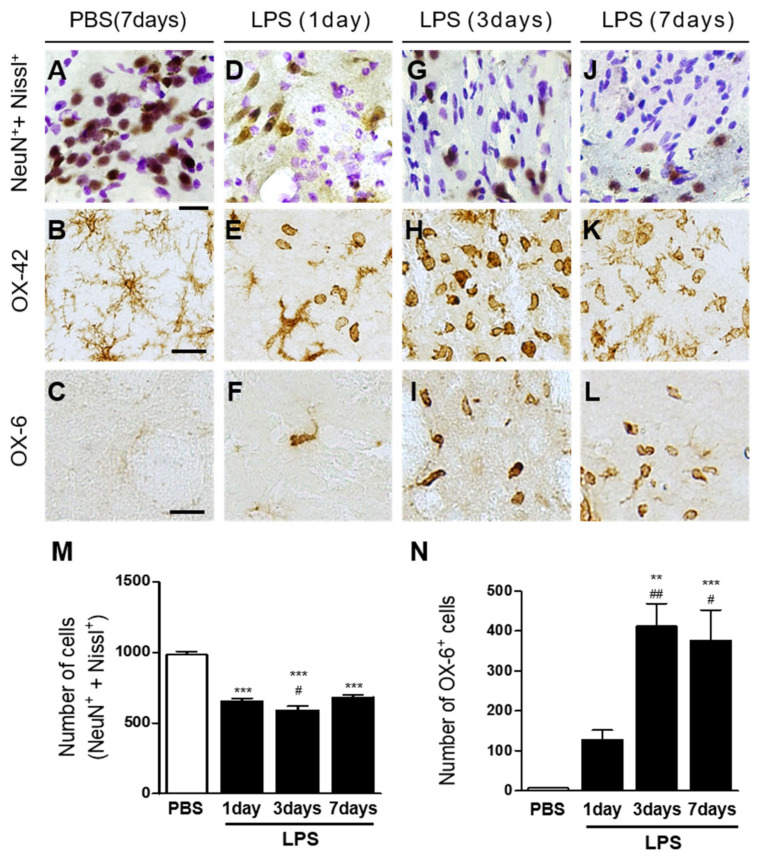
LPS induces neurodegeneration and microglial activation in the rat striatum in vivo. Animals unilaterally received injection of phosphate-buffered saline (PBS) (A–C) as a control or lipopolysaccharide (LPS; (D–L); 5 µg/3 µL) into the rat striatum and were transcardially perfused at indicated time points. The brain tissues were processed for Nissl staining (A,D,G,J) and immunohistochemical staining with NeuN (neuronal nuclei) and Nissl costaining (A,D,G,J) or OX-42 (complement receptor3, CR3; (B,E,H,K)) to identify microglia/macrophages, or OX-6 (major histocompatibility complex class Ⅱ; (C,F,I,L)) to identify activated microglia at 1 day (D–F), 3 days (G–I), and 7 days (J–L) after intrastriatal injection of LPS. Scale bars, 20 µm (A,D,G,J), 25 µm (B,C,E,F,H,I,K,L). (M) Quantification of NeuN+ and Nissl+ cells in the LPS-injected striatum (Total area = 4.6 × 105 μm2). *** p < 0.001, significantly different from PBS (control). Data are presented as the mean ± SEM; n of animals = 4 to 5 in each group, ANOVA with Newman–Keuls analysis. (N) Quantification of OX-6+ cells in the LPS-injected striatum (total area = 4.6 × 105 μm2). ** p < 0.01, significantly different from PBS (control). *** p < 0.001, significantly different from PBS (control). ## p < 0.01, significantly different from LPS 1 day. # p < 0.05, significantly different from LPS 1 day. Data are presented as the mean ± SEM; n of animals = 4 to 5 in each group, ANOVA with Newman–Keuls analysis.

