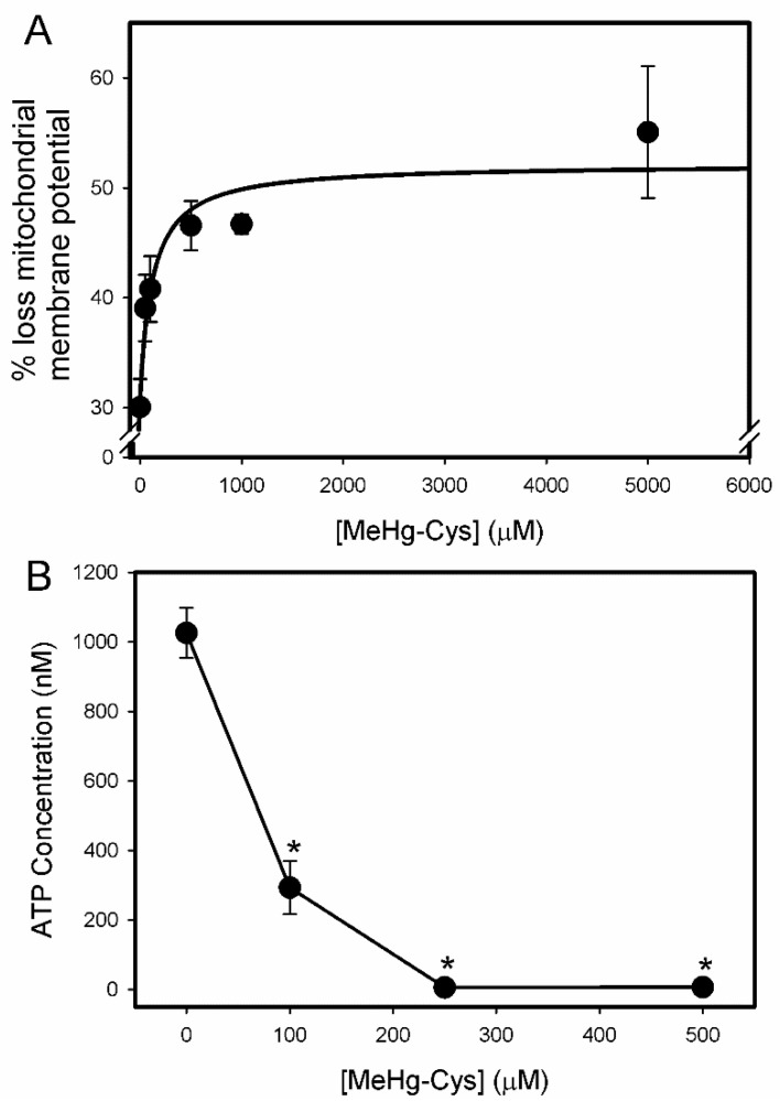
Figure 7.
Mitochondrial disruptions in BeWo cells exposed to MeHg-Cys. BeWo cells were exposed to buffer or various concentrations of MeHg-Cys for 30 min at 37 °C. The loss of mitochondrial membrane potential (A) was measured using fluorescence-activated cell sorting (FACS). Changes in ATP levels were measured under the same exposure conditions (B). Results are presented as mean ± SE. Data represent 3 experiments performed in triplicate. *, significantly different (p < 0.05) from unexposed cells.

