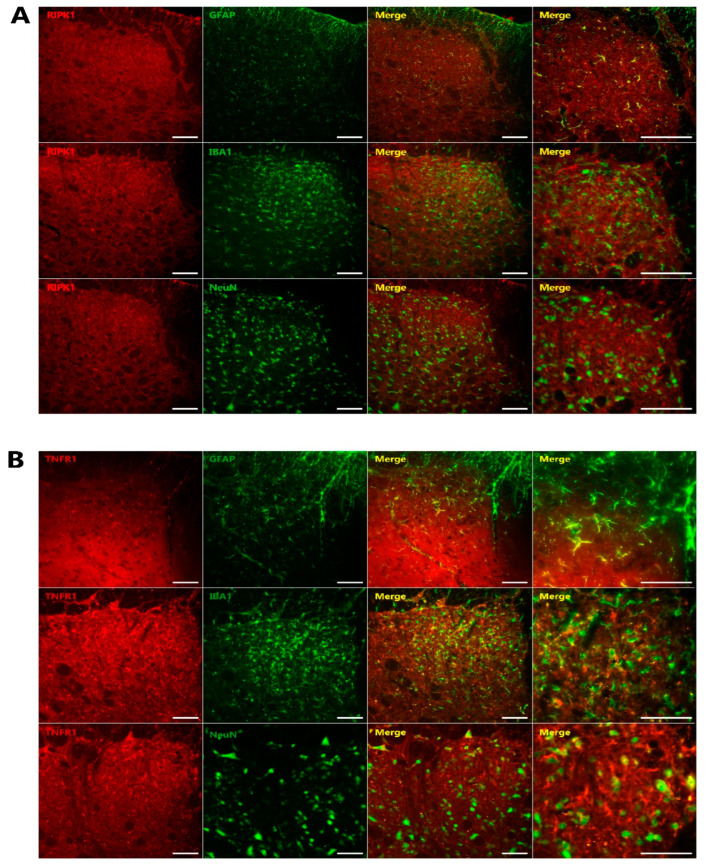
Figure 5.
Characterization of RIPK1 and TNFR1 immunoreactive cells in the TSC after inferior alveolar nerve injury. (A) Double immunofluorescence analysis for RIPK1 (red) and NeuN, a neuronal marker (green); GFAP, an astrocyte marker (green); or IBA1, a microglia marker (green). RIPK1 immunoreactive cells were found to be mainly colocalized with GFAP, an astrocytic marker (green) (n = 6). (B) Double immunofluorescence staining for TNFR1 (red) with NeuN, GFAP, or IBA1 on POD 3. TNFR1 showed colocalization with GFAP (n = 6). Scale bars, 50 µm.

