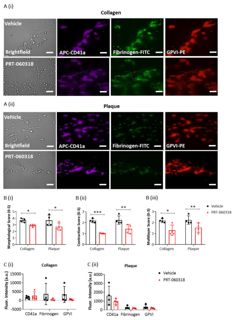
Figure 3.
Platelet activation imaging of thrombi postperfused under condition of Syk inhibition. Recalcified blood samples labelled with APC-αCD41a mAb (purple), FITC-fibrinogen (green) and PE-αGPVI mAb (red) was perfused for 7 min over immobilised collagen or plaque material at room temperature and arterial shear rate (1000 s−1). Post-perfusion for 3 min was with rinse buffer containing vehicle (DMSO) or Syk inhibitor PRT-060318 (10 µM). (A) Representative microscopic images, showing remaining aggregates on collagen (Ai) or plaque (Aii) after 3 min of post-perfusion with vehicle or PRT-060318 (n = 4 donors). (B) Graphs showing scores of thrombus morphology (Bi), thrombus contraction (Bii) and thrombus multilayer (Biii) [16]. (C) Graphs of fluorescence intensity (arbitrary units, a.u.) of platelet aggregates on collagen (Ci) or plaque material (Cii) fixed after 3 min of post-perfusion. Scale bar = 50 µm. Data are shown as mean ± s.d., * p < 0.05, ** p < 0.005, *** p < 0.0005, one-tailed Student’s paired t-test.

