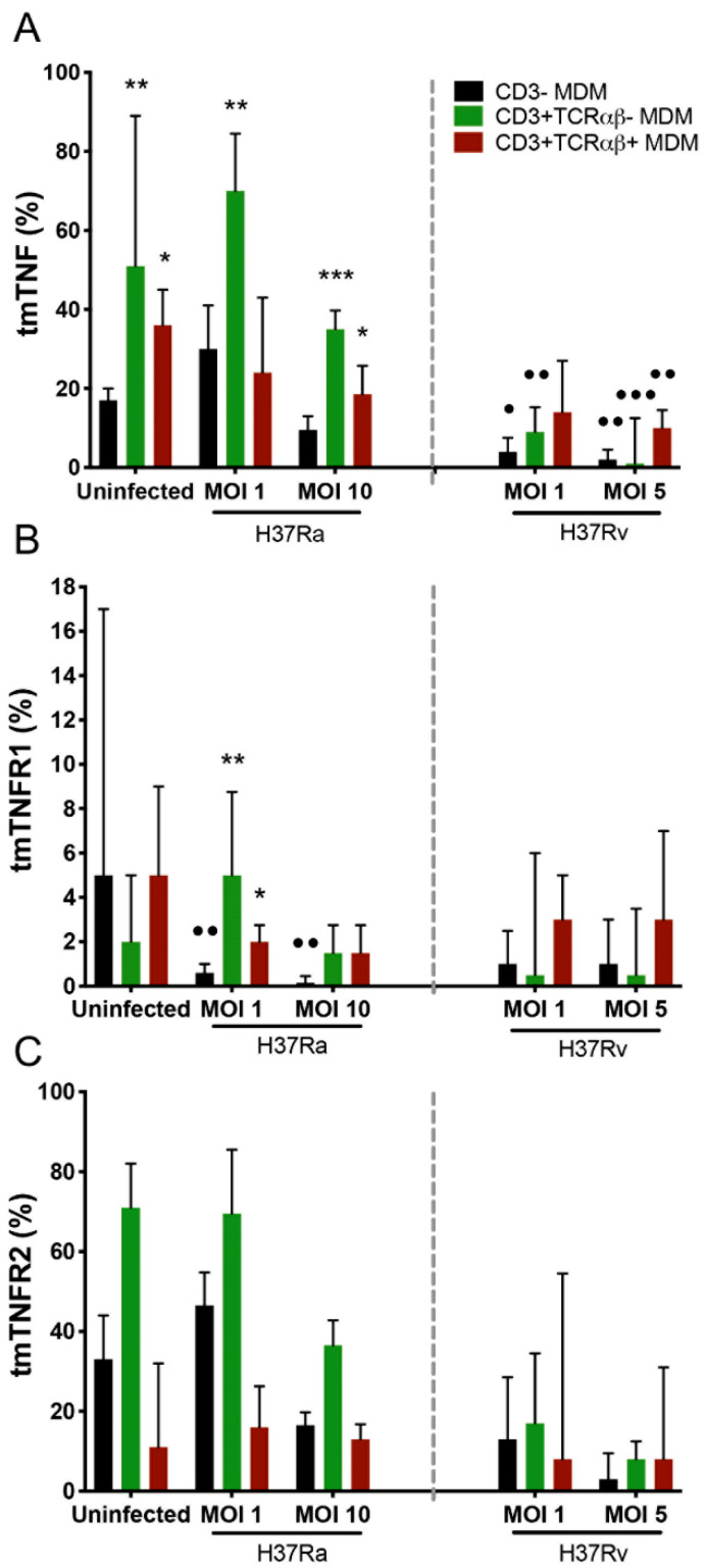
Figure 3.
Expression of tmTNF on myocyte-derived macrophages (MDMs) infected with the Mycobacterium tuberculosis (Mtb) H37Rv virulent strain decreased, whereas that of only tmTNFR1 significantly decreased in MDMs infected with the Mtb H37Ra avirulent strain. After 7 days in culture, 2 × 10⁶ MDMs (per condition) were administered, and at 24 h post-infection, they were recovered for flow cytometric analysis. Frequency of Mtb-infected MDM subpopulations expressing tmTNF (A), tmTNFR1 (B), and tmTNFR2 (C) molecules. Bar graphs show median values and interquartile ranges (IQRs, 10–90) from four independent experiments. Kruskal–Wallis tests followed by Dunn’s post-tests were performed to compare macrophage subpopulations under the same MOI conditions versus the CD3− MDM subpopulation, * p < 0.05, ** p < 0.01, *** p < 0.001, and, to compare the infected macrophage subpopulation versus its counterpart under uninfected conditions, • p < 0.05, •• p < 0.01, ••• p < 0.001.

