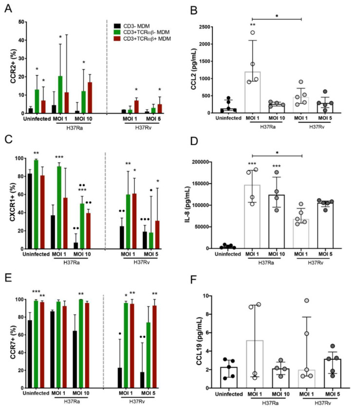
Figure 5.
Chemokine receptors and their ligands implicated in the host inflammatory response are differentially expressed in Mycobacterium tuberculosis (Mtb) H37Ra- or H37Rv-infected CD3− and CD3+ monocyte-derived macrophages (MDMs). After 7 days in culture, 2 × 10⁶ MDMs (per condition) were infected, and at 24 h post-infection, MDMs were recovered for flow cytometric analysis, and the supernatants were recovered for ELISA. (A) CCR2, (C) CXCR1, and (E) CCR7 chemokine receptors were quantified by flow cytometry. Soluble levels of the (B) CCL2, (D) IL-8, and (F) CCL19 chemokines were assessed in culture supernatants by ELISA. Bar graphs in A, C, and E show median values and interquartile ranges (IQRs, 10–90) from four independent experiments; scatter plots with bar in B, D, and F show median values and IQRs from five independent experiments. Kruskal–Wallis tests followed by Dunn’s post-tests with bar were performed to compare macrophage subpopulations under the same MOI conditions versus the CD3− MDM subpopulation, * p < 0.05, ** p < 0.01, *** p < 0.001, and to compare the same macrophage subpopulation in a different condition of culture, • p < 0.05, •• p < 0.01, ••• p < 0.001. In addition, for the analysis of soluble chemokine levels, the Kruskal–Wallis test followed by Dunn’s post-test was performed to compare infected versus uninfected MDMs or between groups (when indicated), * p < 0.05, ** p < 0.01, *** p < 0.001.

