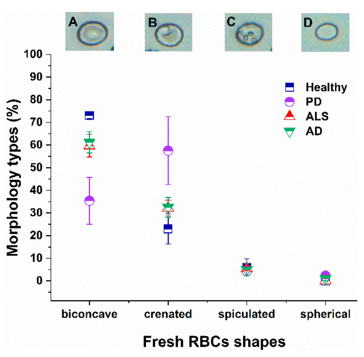
Figure 1.
Optical images of the four different RBCs shapes (biconcave (A), crenate (B), spiculated (C) and spherocytic (D)). Distribution of RBC morphological types (in percentage) in fresh healthy (navy squares), PD (violet circles), ALS (red triangles) and AD (green inverted triangles) cells. Mean values and SD; p values (NDD vs. healthy control group): p < 0.01 for biconcave shape of PD, AD and ALS, and for crenate shape of PD cells; and p < 0.05 for crenate ALS and AD cells.

