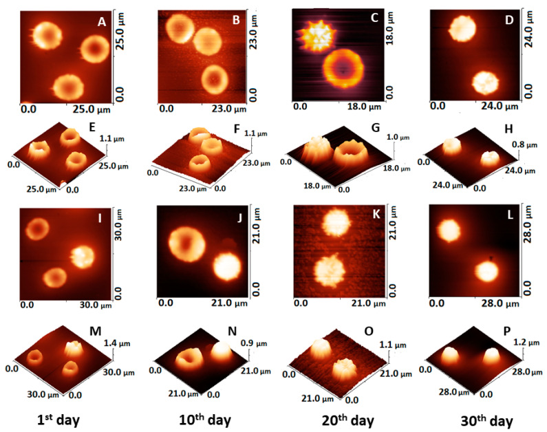
Figure 4.
AFM images taken on smears of fresh and aged RBCs from healthy and PD donors on glass support, the scanned area is given on each image. 2D and 3D images of healthy cells: fresh (A,E), 10-day-aged (B,F), 20-day-aged (C,G) and 30-day-aged (D,H), and of PD cells: fresh (I,M), 10-day-aged (J,N), 20-day-aged (K,O) and 30-day-aged (L,P).

