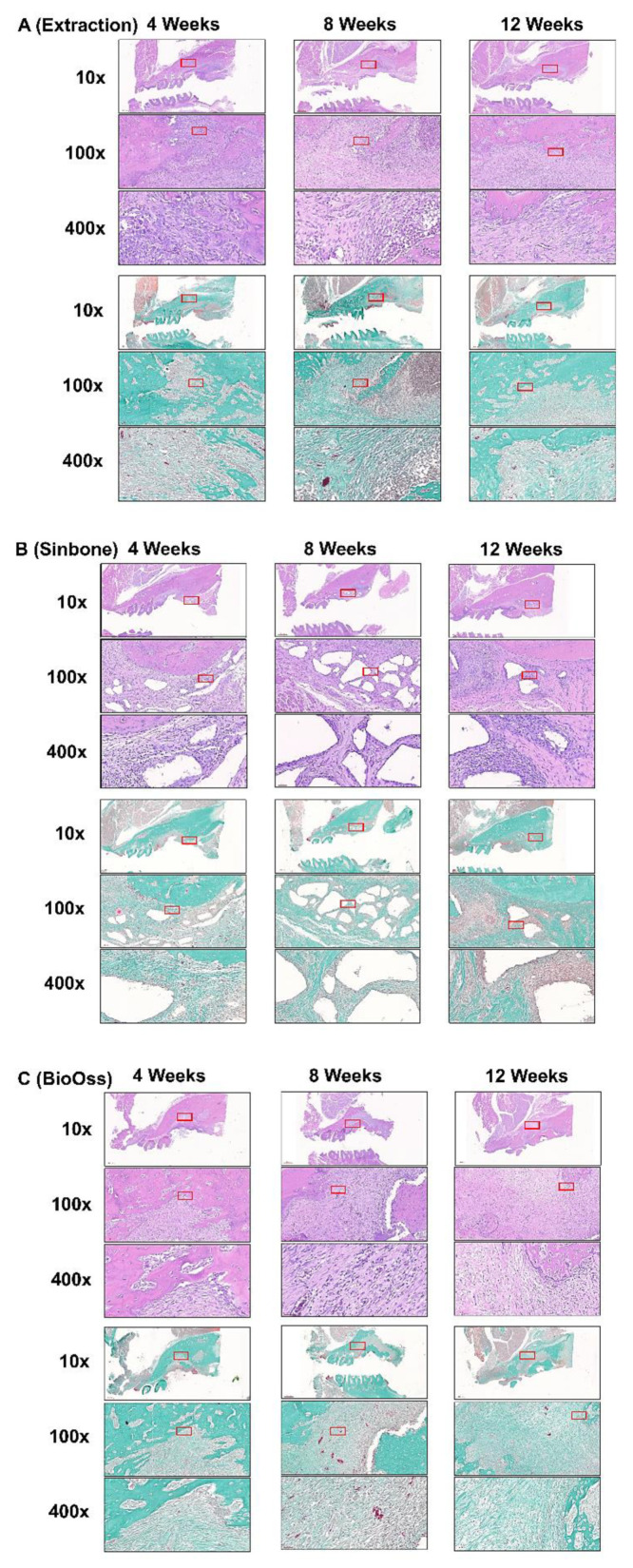
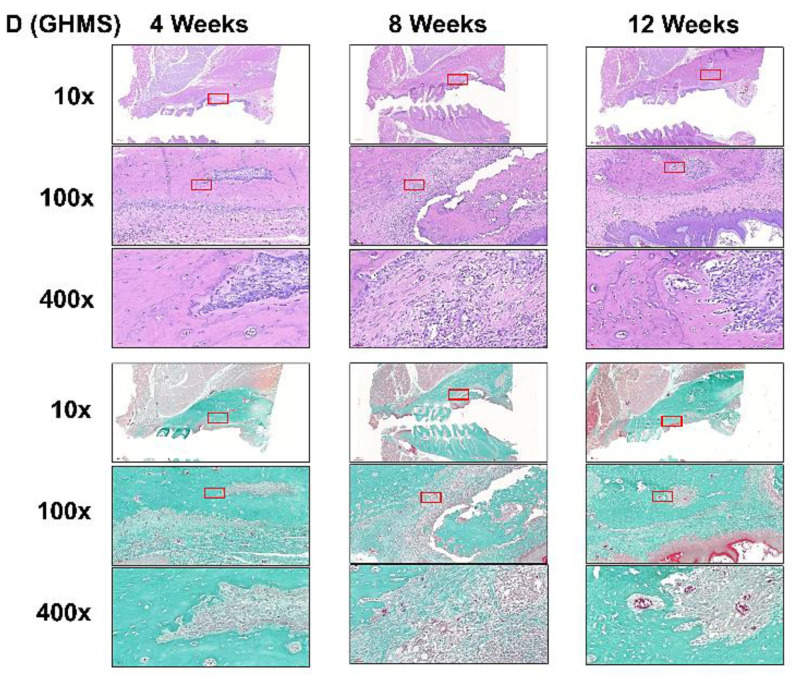
Figure 4.
Representative histological sections of the maxillary first molar (M1) to third molar (M3) region of rats after M1 (A) extraction or grafting with (B) Sinbone, (C) Bio-Oss Collagen®, or (D) gelatin/nano-hydroxyapatite/metformin (GHMS) by hematoxylin and eosin (H&E) and Goldner’s Trichrome staining at 4, 8, and 12 weeks. At the 12th week after extraction, new bone growth was less evident in the extraction-only group. The defects grafted with Sinbone granules showed a substantial amount of graft material remained in the defect site. Bio-Oss Collagen® showed some bone regeneration in the defect site. In contrast, well-developed new bone and the presence of haversian canals were found in the GHMS sections. In addition, there was no detectable fibrous tissue invasion or inflammatory cells infiltration in the GHMS experimental group. Goldner’s trichrome staining shows significantly less new bone formation in the extraction-only group, whereas immature bone formation and soft tissue invasion were observed in Sinbone. In the GHMS samples, there was a well-developed new bone, and angiogenesis were found. The red box is the position of magnification.


