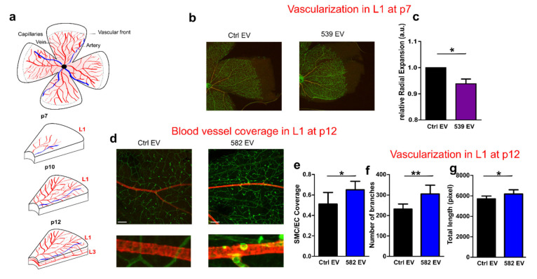
Figure 5.
miR-539 EVs and miR-582 EVs participate in vascular remodeling of the retina. (a) Cartoon illustrating the development of the superficial (L1), intermediate (L2), and deep vascular plexus (L3). (b) Left: retinal flat mount representation. For (b–g), EVs were purified from ECs transfected with pre-miR-539 (539 EV) or a pre-miR-Control (Ctrl EV) or from SMCs transfected with pre-miR-582 (582 EV) on a pre-miR-control (Ctrl EV) (b) Isolectin-B4 staining on Postnatal Day 7 retinas from pups that were injected at Postnatal Day 1 with EVs. Scale bar: 25 µm. (f–h). The histograms represent the quantification of the radial expansion from the optic nerve to the vascular front (c), N = 8 eyes, two independent experiments. (d) Confocal images of isolectin-B4/α -SMA staining on postnatal Day 12 retinas from pups that were injected at Postnatal Day 7 with miR-582 EVs in the superficial layer (L1). Scale bar: 200 µm. (e–g). The histograms represent the quantification of the coverage of ECs by SMCs (Spearman’s rank correlation value) (e), the number of branches (f), and the total length of isolectin+ vessels (g) * p < 0.05, ** p = 0.01.

