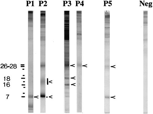
FIG. 1.
Echinococcus Western Blot IgG. Five immunoblot patterns (P1 through P5) are obtained with cystic and alveolar echinococcosis sera. Most of the significant bands are indicated by arrows. Molecular sizes (in kilodaltons) are indicated on the left. Neg, results for negative-control serum. Reprinted by courtesy of LDBIO Diagnostics.

