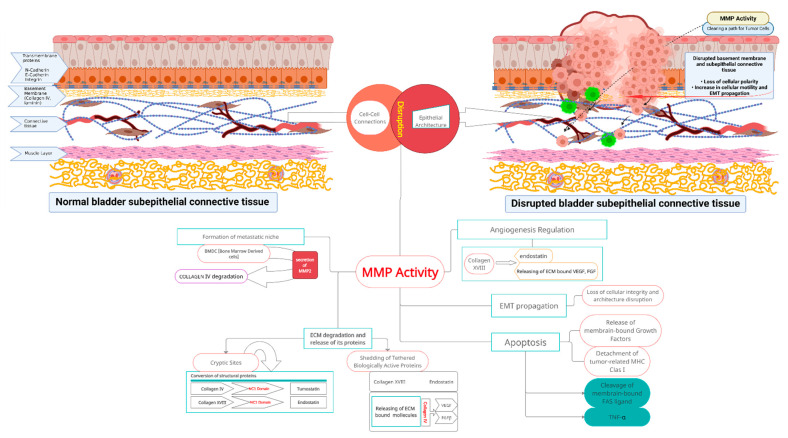
Figure 1.
MMP activity and related molecular events during bladder cancer propagation. Upper left part of the diagram: Normal bladder wall structure. Correct epithelial architecture and polarity provided by the indicated adhesion molecules, and with a normal basement membrane. Layered self-assembled collagen IV and laminin networks blend intact basement membrane (yellow). Regular structure of underlying connective and muscle tissues. Intact layered structure of bladder wall is crucial in proper bladder physiology. Upper right part of the diagram: Gradual bladder wall disruption related to aberrant MMP activity. Cleavage of intercellular junctions, along with basement membrane remodeling and further loss of polarity of the urothelium, increased cell motility with propagation of epithelial-to-mesenchymal transition. Cancer cells traverse degraded supporting barrier (formerly continuous basement membrane). Center of the diagram: Degradation of extracellular matrix mediated by matrix metalloproteinases 2 and 9 released by cancer cells, and accompanying cells from tumor-associated microenvironment cleave components of the basement membrane and underlying connective tissue, and generate new derivative molecules. As a result, extracellular matrix structural proteins and matrix-bound latent signaling molecules are converted into biologically active signaling molecules that are converted into active mediators. Activity of those products (products listed in the following segments of central part of the scheme) results in further cancer cells chemoattraction, proliferation, infiltration of deep tissues, aberrant neo-angiogenesis, inhibition of apoptosis in cancer cells, and formation of cancer niches. These molecular events contribute to bladder cancer progression from non-muscle to muscle-invasive tumors. MMP actions that take place after MMP activation are explained in the body text.

