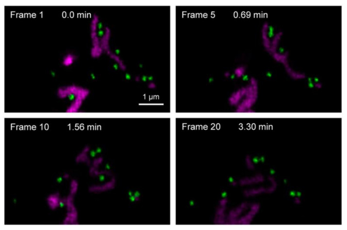
Figure 4.
Two-color STED super-resolution imaging of living HeLa cells. The lipid droplets (green) and mitochondria (purple) were labeled with Lipi-DSB and TMRM, respectively (scale bar = 1 μm); the STED images were recorded under two excitation lasers (470 nm for Lipi-DSB, 550 nm for TMRM) and one STED laser (660 nm CW-STED, 40 MW cm−2). Reprinted (adapted) with permission from Zhou, R.; Wang, C.; Liang, X.; Liu, F.; Yan, X.; Liu, X.; Sun, P.; Zhang, H.; Wang, Y.; Lu, G. Stimulated Emission Depletion (STED) Super-Resolution Imaging with an Advanced Organic Fluorescent Probe: Visualizing the Cellular Lipid Droplets at the Unprecedented Nanoscale Resolution. ACS Mater. Lett. 2021, 3, 516–524, Copyright 2021 American Chemical Society.

