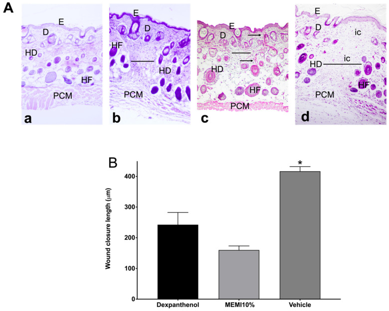
Figure 6.
Histological analyses of MEMI. (A) Histological sections of skin wounds on mice. Control group (Dexpanthenol) showed a thicker epidermis than that of MEMI 10% group on wound´s contraction. Representative photographs of the wound’s architecture on the 14th day: normal skin (a); dexpanthenol group (b); MEMI 10% group (c); surgical gel vehicle (d); epidermis (E); dermis (D); hypodermis (HD); hair follicle (HF); panniculus carnosus muscle (PCM); inflammatory cells (ic); measurement wound (-); collagen fibers (←). Tissues were stained with H&E and visualized at 10× magnification. (B) Reduction in wound on 14th day of treatment. Results were expressed as the mean ± S.D. The analysis of the data was conducted using two-way ANOVA with a Tukey multiple comparison post hoc test. * p < 0.05 compared to dexpanthenol and MEMI 10% groups.

