
Figure 3. p21high cells accumulate in various tissues with aging.
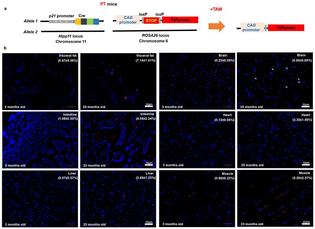
(a) Schematic of PT mice. (b) Representative micrographs of 6 tissues in young and old PT (male and female) mice. Red: tdTomato, Blue: DAPI. The percentages of tdTomato+ cells are shown as means ± s.e.m. Experiments were repeated in 3 mice.