
Figure 4. p21high cells can be induced by aging, chemotherapy, and obesity.
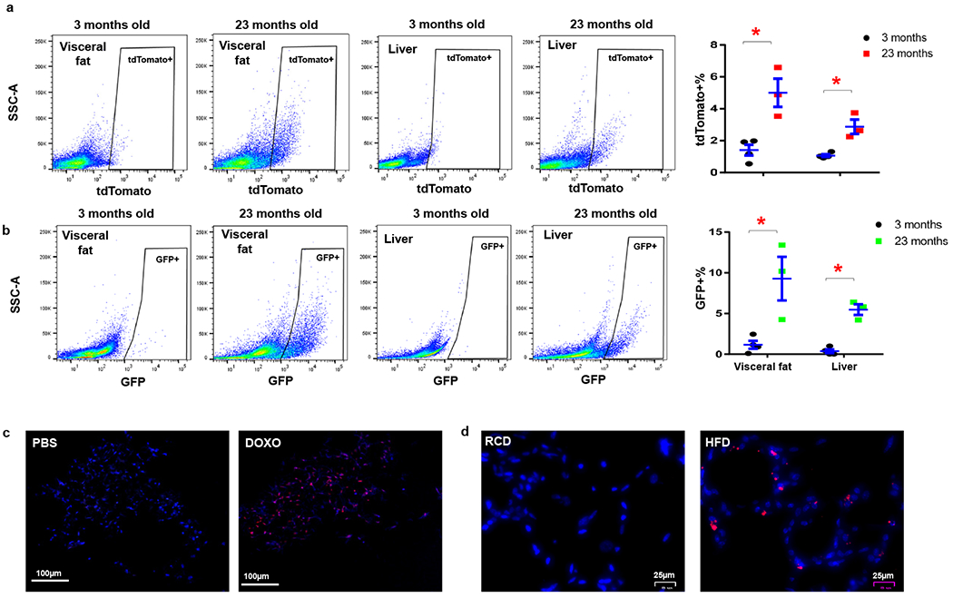
(a) Flow cytometry analysis and proportion of tdTomato+ p21high cells in 3-month-old (n=4) and 23-month-old (n=3) PT mice. * p=0.008 for fat, p=0.005 for liver; (b) Flow cytometry analysis and proportion of GFP+ p21high cells in 3-month-old (n=4) and 23-month-old (n=3) PT mice. Results are shown as means ± s.e.m. * p=0.017 for fat, p<0.001 for liver; two-tailed, unpaired Student’s t-test. (c) Representative micrographs of visceral fat in 3-month-old PT mice treated with PBS or DOXO. Red: tdTomato, Blue: DAPI. Experiments were repeated in 3 mice. (d) Representative micrographs of visceral fat in 5-month-old PT mice fed with RCD or HFD for 2 months. Red: tdTomato, Blue: DAPI. Experiments were repeated in 4 mice.