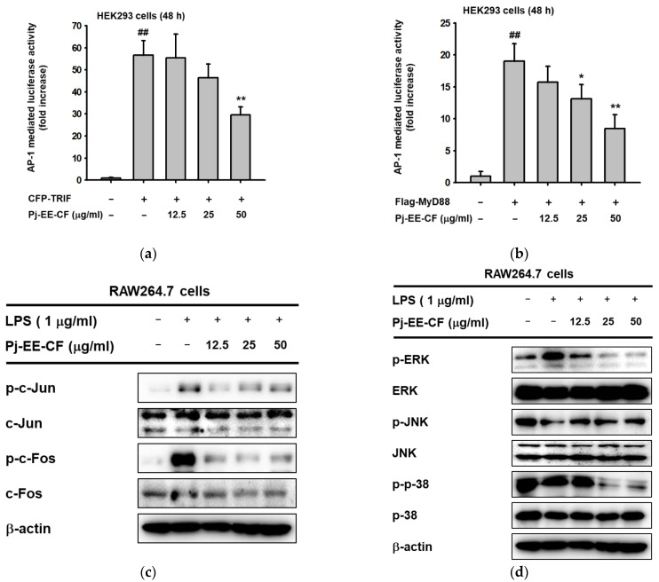
Figure 3.
Effect of Pj-EE-CF on the transcriptional activation of AP-1 signaling and the upstream signaling molecules of AP-1 activation. (a,b) HEK293 cells were co-transfected with AP-1-Luc and β-gal (0.8 μg), as well as TRIF and MyD88 for 48 h in the presence or absence of Pj-EE-CF (12.5, 25, and 50 μg/mL), which were then examined using a luminometer. Results are expressed as mean ± standard deviation (n = 4). ## p < 0.01 compared to normal group, * p < 0.05 and ** p < 0.01 compared to control group (LPS alone) by one-way ANOVA. (c) The phospho- and total forms of AP-1 subunits, c-Jun and c-Fos, from whole-cell lysates from LPS-treated RAW264.7 cells in the presence or absence of Pj-EE-CF (12.5, 25, and 50 μg/mL) were determined by immunoblot analysis. (d) RAW264.7 cells were pretreated with Pj-EE-CF (12.5, 25, and 50 μg/mL) for 30 min, followed by the presence or absence of LPS. The phosphorylated and total protein levels of ERK, JNK, and p38 were assessed by immunoblot analysis. β-actin was utilized as a control protein.

