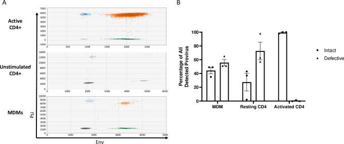
Fig 1. Acute HIV-1 infection generates defective proviruses in MDMs and resting CD4+ T cells.
(A) Representative raw IPDA data of (top) activated CD4+ T cells, (middle) unstimulated CD4+ T cells, and (bottom) monocyte-derived macrophages (MDMs) infected with HIV-1. Droplets are color coded based on manual gating of positive and negative probe signals (gray = empty or double mutation/deletion droplets, blue = droplets single positive for psi, green = single positive env droplets, orange = double positive psi and env intact proviral droplets). A parallel reaction to detect the host cell gene RPP30 was used as a correction for DNA shearing. (B) Percentages of intact and defective HIV genomes detected in MDMs and CD4+ T cells following HIV-1 infection. MDMs were infected with HIV-1NL4-3-BaL for 48 hours prior to DNA isolation. Resting and anti-CD3/CD-28 bead activated CD4+ T cells were infected with HIV-1NL4-3-VSVg for 72 hours prior to DNA isolation. Data represents three independent infections using cells generated from three different donors. Resting and activated CD4+ T cell sample data are participant matched.

