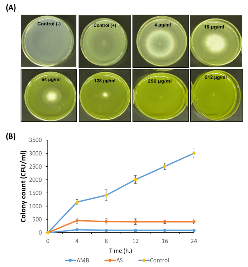
Figure 3.
Antimicrobial potential of AS-AgNPs. (A) A. fumigatus spores were added to the center of PDA plates containing different concentrations of AS-AgNPs (4, 16, 64, 128, 256, and 512 μg/mL). The positive control (amphotericin B) was treated with DMSO (less than 1%), and the negative control was only PDA without fungus. The MIC was identified as the lowest concentration of AS-AgNPs that does not show any growth of fungal colonies on the agar plates. At concentrations of more than 128 μg/mL, there were no colonies visible on the plates. (B) Time–kill curves of A. fumigatus following exposure to AS-AgNPs and amphotericin B. Values are expressed as means ± SD.

