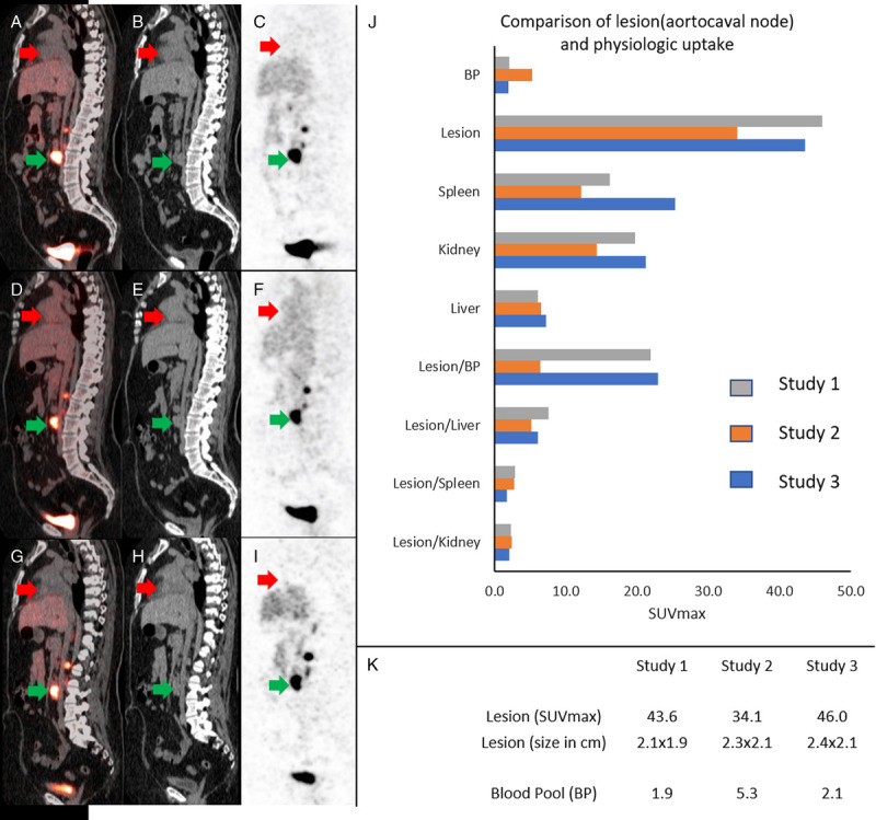
FIGURE 2.

Sagittal images from serial 68Ga-DOTATATE PET/CT show inverse relationship between nodal disease and blood pool uptake. Initial time point 68Ga-DOTATATE sagittal fused (A), CT (B), and PET (C) demonstrate low blood pool uptake (red arrows; SUVmax, 1.9) and high 68Ga-DOTATATE uptake in a dominant aortocaval lymph node (green arrows; SUVmax, 43.6). Sagittal fused (D), CT (E), and PET (F) of 2 months' follow-up 68Ga-DOTATATE show high blood pool SUV values (red arrows; SUVmax, 5.3) and corresponding drop in 68Ga-DOTATATE uptake in the aortocaval lymph node (green arrows; SUVmax, 34.1), which returned to baseline values on subsequent 68Ga-DOTATATE imaging with fused (G), CT (H), and PET (I) with low blood pool uptake (red arrows; SUVmax, 2.1) and high lymph node uptake (green arrows; SUVmax, 46). In this case, the lesion and blood pool uptake showed greater variability than in liver, spleen, and kidney (J). There was no substantial change in lymph node sizes between the 3 studies on CT images (K).
