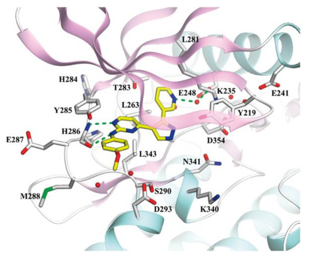
Figure 81.

X-ray crystal structure of lead compound 75 in R206H mutant ALK2. White stick model: residues forming ATP binding pockets; Yellow stick model: lead compound; Green dashed lines: the hydrogen bonds between lead compound and active site; Red sphere: water molecules [88].
