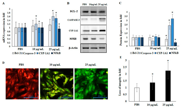
Figure 5.
Effects of EO from A. lanatum on the mRNA and protein markers of HepG2 liver cancer cells. The effects of EO on the inhibition of apoptotic and angiogenic markers were evaluated by real-time PCR. (A) HepG2 liver cancer cells were supplemented with EO (10 and 25 μg/mL). The mRNA of apoptotic and angiogenic markers that was altered in EO-treated cells was quantified using quantitative real-time PCR. GAPDH was used as an internal mRNA control. (B,C) Alterations in the status of metastasis-associated proteins in response to EO supplementation were inspected using Western blot. HepG2 cells were supplemented with EO (10 and 25 μg/mL) for 24 h. β-actin was utilized as a control. (D,E) The mitochondrial membrane potential (MMP) was estimated for in EO-treated (10 and 25 μg/mL) HepG2 cell lines after an incubation period of 24 h. The mitochondrial membrane integrity was analyzed using the emission of green fluorescent by ImageJ software. The experimental data are shown as the mean ± SD of triplicate values; * p < 0.05 when evaluated against control (PBS; phosphate-buffered saline).

