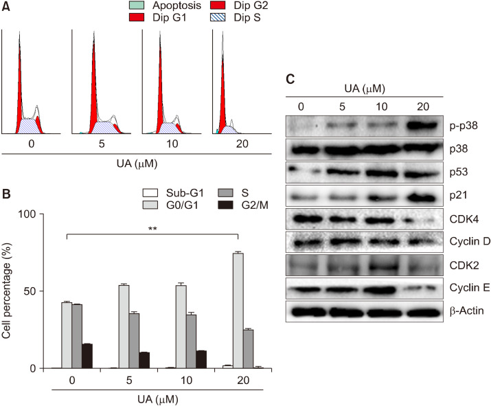
Fig. 2.
Effect of ursolic acid (UA) on cell cycle progression in MCF-7 cells. (A) Flow cytometry analyses of the distribution of the stages of the cell cycle in MCF-7 cells stained with propidium iodide. MCF-7 cells were treated with UA (0, 5, 10, and 20 μM) for 48 h. (B) The percentage of cells accumulated at G0/G1, S, and G2/M phases of the cell cycle depending on the cell type. Results are expressed as mean±SD. Statistical differences were analyzed using Student’s t-test (**P<0.01 vs. control). (C) Expression of the G0/G1-related proteins p-p38, p38, p53, p21, CDK4, cyclin D, CDK2, and cyclin E, examined by Western blotting.

