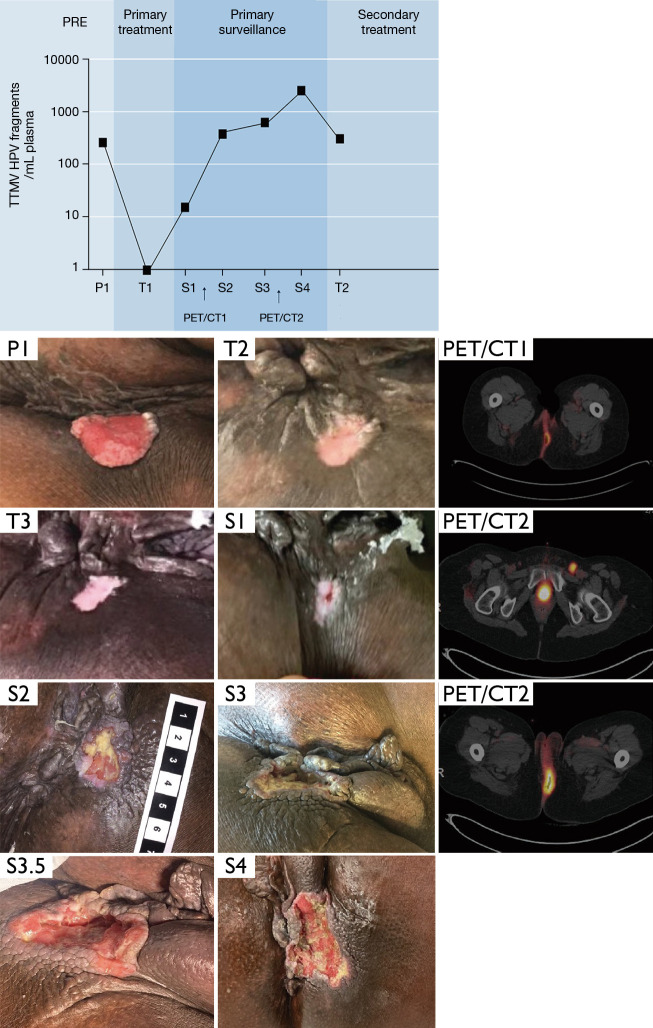
Figure 1.
A 48-year-old woman with several medical co-morbidities presented with a T2N0M0 HPV-associated anal cancer. Plasma TTMV-HPV DNA values and photographs are shown at the indicated treatment time intervals: pre-treatment (P), during treatment (T), and post-treatment surveillance (S). At initial presentation, there was a 2.5 cm × 3.5 cm left lateral anal margin mass without involvement of the anal canal. Examination improved by week 4 of treatment (T1) at which time TTMV-HPV16 DNA was undetectable. Only a superficial ulceration of the skin was observed at week 8 (T2) and week 12 of treatment (T3). Two months after therapy, she presented with pain and ulceration (S1), and TTMV-HPV16 DNA was detectable. Biopsy confirmed local recurrence. PET/CT showed hypermetabolic cutaneous activity suspicious for local tumor recurrence (PET/CT1). With further local progression at the primary site (S2, S3, S3.5) and progression on the PET/CT to involve ipsilateral pelvic and groin lymph nodes (PET/CT2), she underwent salvage chemoRT (S4), with a subsequent TTMV-HPV16 DNA drawn in her last week of therapy (T2). TTMV-HPV DNA, tumor-tissue-modified HPV DNA.

