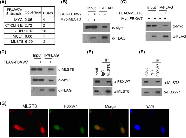
FIGURE 5.

FBXW7 directly interacts with MLST8. A, Quantitative mass spectrometry using FBXW7 knockout (KO) HCT116 cells. B and C, 293T cells were co‐transfected with Myc‐MLST8 and FLAG‐FBXW7 constructs. D, Western blot and co‐IP samples of anti‐FLAG antibody obtained from 293T cells infected with expressing FLAG‐FBXW7 or control. E and F, After treatment with 20 μm MG132 for 4 h, 786‐O cell lysates were prepared for Co‐IP with anti‐MLST8 antibody and WB analyses with indicated antibodies. G, Confocal microscopy of 786‐O cells stained with MLST8 and FBXW7 antibodies. DAPI represents nuclear staining. Red, green, and blue channel images were captured by Olympus. Maximum projection images are shown, original magnification ×40
