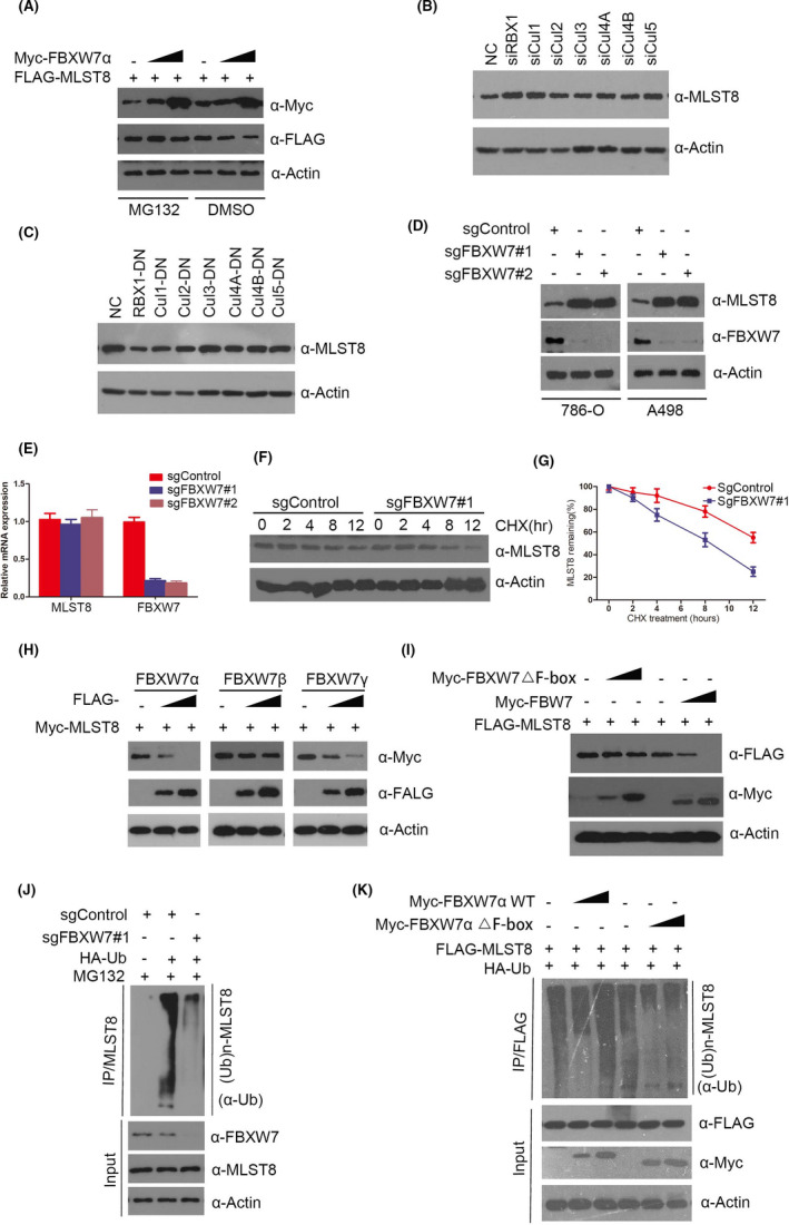
FIGURE 6.

MLST8 is degraded and ubiquitination by tumor suppressor FBXW7. A, Western blotting analysis of WCLs of 293T cells transfected with the indicated constructs. B, The expressions of Cul1, Cul2, Cul3, Cul4A, Cul4B, Cul5, and RBX1 were reduced by transient transfection siRNA in 786‐O cells. Protein samples were collected 48 h after transfection, and the reduction effect and expression of MLST8 were detected by Western blot. C, In 786‐O cells, Cul1, Cul2, Cul3, Cul4A, Cul4B, and Cul5 mutants with the fLag label were overexpressed. Protein samples were collected 48 h after transfection, and the overexpression effect and expression level of MLST8 were detected by Western blot. D, Western blot of indicated proteins in WCLs from 786‐O and A498 cells with FBXW7 knockout through CRISPR/Cas9 methods. E, RT‐qPCR measurement of MLST8 mRNA expression in 786‐O cells infected with lentivirus expressing FBXW7‐specific sgRNA or NC; RT‐qPCR measurement of MLST8 mRNA expression in parental and FBXW7‐KO 786‐O cells. Data are shown as means ± SD (n = 3). F and G, Western blot of indicated proteins in WCLs of 786‐O cells infected with lentivirus expressing FBXW7‐specific sgRNA or NC for 48 h and then treated with 50 μg/mL cycloheximide (CHX) and harvested at different time points. H, 786‐O cells were transfected with the indicated plasmids for 24 h, followed by Western blotting analysis. I, 786‐O cells were transfected with the indicated plasmids for 24 h, followed by Western blotting analysis. J, Western blot of indicated proteins in WCLs from 786‐O with FBXW7 knockout through CRISPR/Cas9 methods, followed by treatment with 20 μM MG132 for 8 h. K, 786‐O cells were transfected with the indicated plasmids for 16 h, followed by treatment with 20 μM MG132 for 8 h
