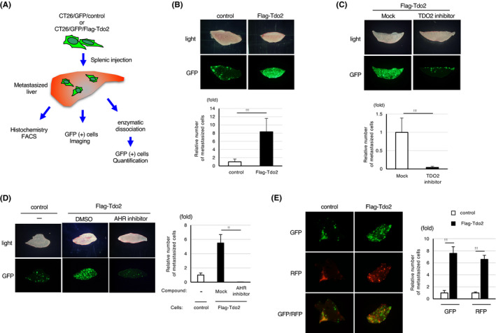
FIGURE 2.

Tdo2 expression promotes liver metastasis in an Ahr‐dependent manner. A, An experimental scheme of the liver metastasis analyses. B, Promotion of liver metastasis by colon cancer cells induced by Tdo2 expression. Top: Representative stereo microscopy and GFP images of the liver at 7 d after splenic injection of CT26‐derived cells. Bottom: Relative numbers of liver‐metastasized GFP‐positive cells. C, Inhibition of Tdo2‐induced liver metastasis by daily intraperitoneal injection of a Tdo2 inhibitor. Top: Representative GFP images of liver metastases. Bottom: Relative numbers of liver‐metastasized GFP‐positive cells. D, Inhibition of Tdo2‐mediated liver metastasis by daily intraperitoneal injection of an AHR inhibitor. Left: Representative GFP images of liver metastases. Right: Relative numbers of metastasized GFP‐positive cells. E, Promotion of liver metastasis by coinjected Tdo2‐expressing cells. Left: Representative fluorescent images of liver metastases. Right: Relative numbers of liver‐metastasized cells. Values represent the mean ± SD; **P < .01, ***P < .001 (two‐sided Student's t‐test)
