FIGURE 3.

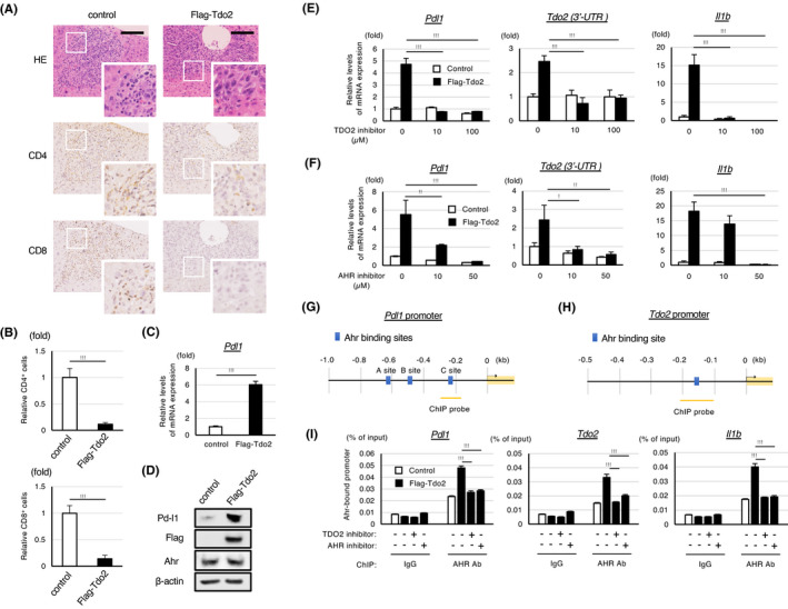
The induction of the TDO2‐kynurenine‐AHR pathway transactivates Pd‐l1 and compromises immune responses in metastatic liver. A, H&E staining and immunohistochemical analyses of metastasized livers at 7 d after the splenic injection of the CT26/GFP cells expressing the indicated construct. Images acquired at a higher magnification are also shown. Scale bar: 100 µm. B, Relative numbers of infiltrating CD4+ and CD8+ cells in the indicated tumors shown in (A). C, qPCR analysis of Pdl1 expression in CT26 cells expressing Flag‐Tdo2 and control cells. D, Western blot analyses of the expression of the indicated proteins in the cells shown in (C). E, F, qPCR analyses of the expression of the indicated genes in the cells shown in (C). The cells were treated with the indicated amounts of a TDO2 or AHR inhibitor for 48 h before the analyses. For quantification of endogenous Tdo2 expression, 3’‐UTR of Tdo2 was evaluated. G, H, Schematic representations of the predicted Ahr binding sites in the Pdl1 and Tdo2 promotors. I, ChIP analyses of AHR binding to the indicated promotor region in CT26/GFP/Flag‐Tdo2 cells or control cells. The cells were treated with the TDO2 or AHR inhibitor for 48 h or mock treated. Enrichment over input (% input) was measured by qPCR (n = 3). As for the ChIP analyses of the Pdl1 promoter, the results of the AHR binding for the C site shown in (G) are shown (the binding to the other sites was not detected). Values represent the mean ± SD; *P < .05, **P < .01, ***P < .001
