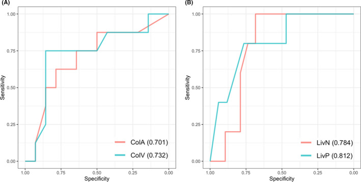
FIGURE 4.

Receiver operating characteristic plots for machine learning prediction for high or low tumor mutational burden in the primary (A) and metastatic (B) lesions. ColA and ColV, computed tomography (CT) of arterial and venous phases for the primary colon lesion. LivN and LivP, CT of non‐contrast enhancement and portal phase for the metastatic liver lesion. Area under the curve is shown in parentheses
