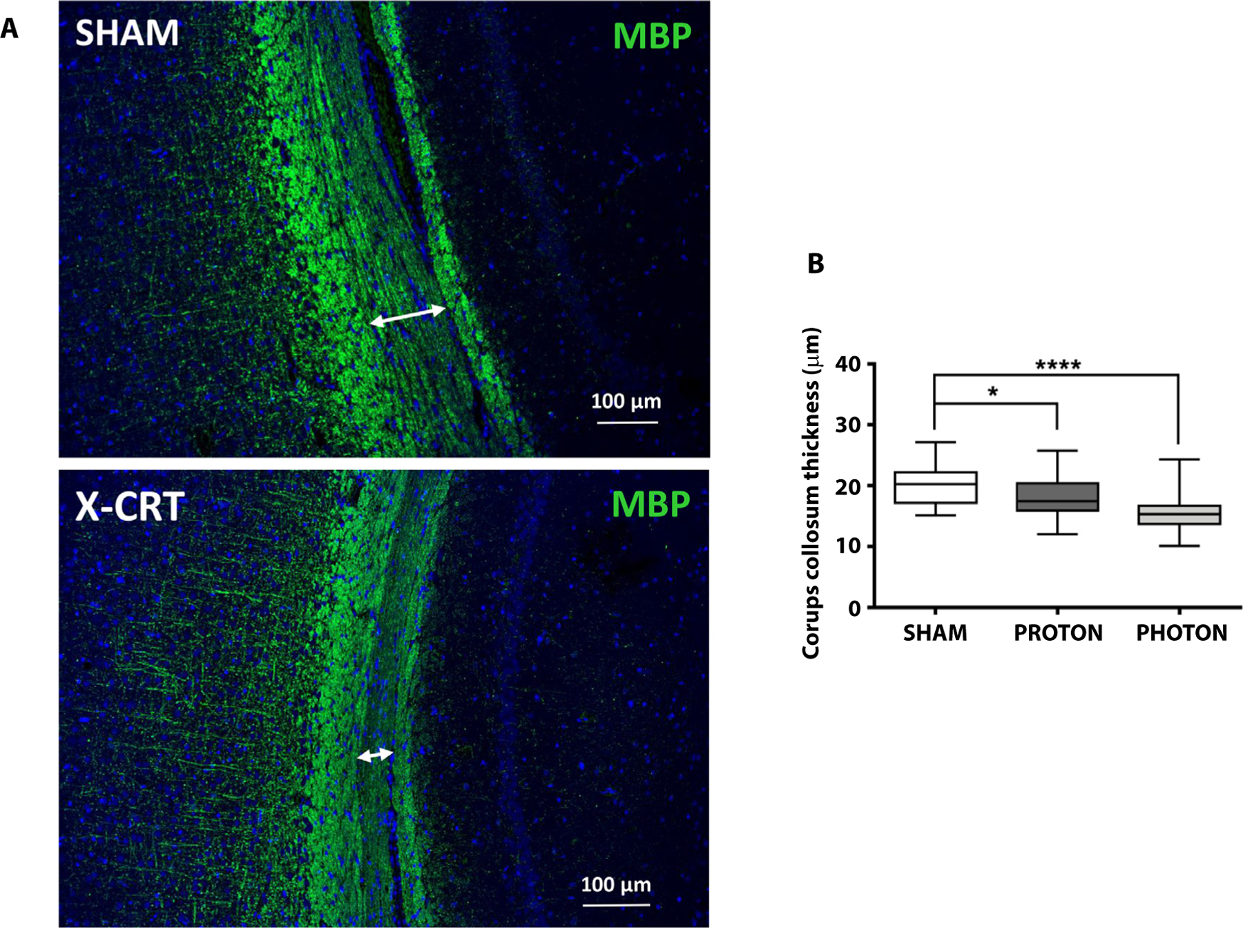
Fig. 6.

CRT brain at 12 months post-CRT showed significant myelin thinning. (A) CRT brain sections immunostained for myelin basic protein (MBP) in sham (top) vs X-CRT (lower), and (B) quantification of the corpus callosum thickness. Data are shown as box-and-whisker plot: box extends from the 25th to 75th percentiles, median value marked by line and whiskers extend from min to max, n = 4–5/group. *P < .05 and ****P < .0001. Scale bar represents 100 μm. Abbreviations: CRT = cranial radiation therapy; X-CRT = photon cranial radiation therapy.
