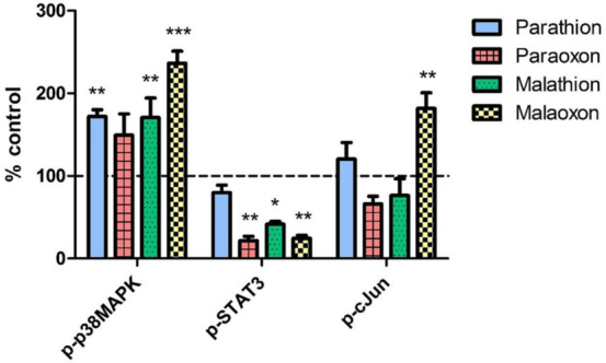
Fig. 6.

Phosphorylation of signaling pathways after OP exposure. PCLS were exposed to 1000 µmol/L of parathion, paraoxon, malathion or malaoxon for 8 h. Intracellular levels of the phosphorylated proteins p-p38MAPK, p-STAT3 and p-c-Jun were evaluated using a Bioplex system. Data are calculated as % of control (indicated by dashed line) and are shown as mean ± SEM. Asterisks indicate significant differences to the control (*p < 0.05; n = 4 samples from four different animals)
