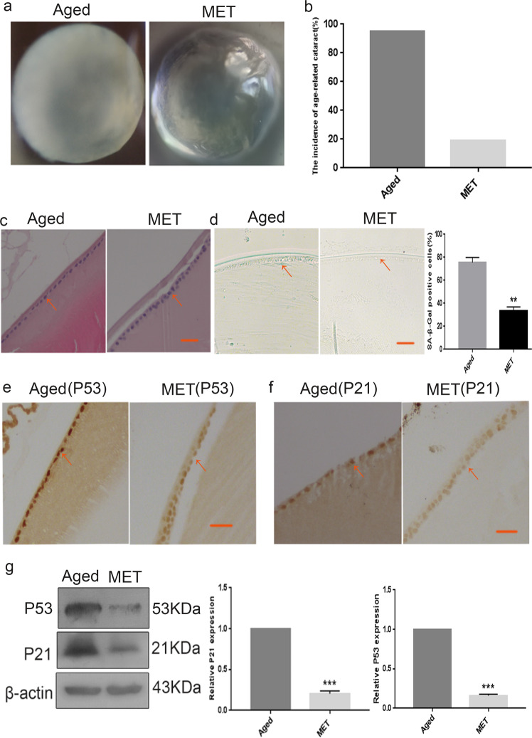
Fig. 2. Chronic low-dose MET administration served the transparency of lens and alleviated the senescence of lens epithelial cells.
a Representative images of morphological observations of lens in the Aged (left) and the MET (right). b The cataract incidence in the Aged (left) and the MET (right). c The lens of the Aged (left) and the MET (right) were analysed by H&E staining. d Representative images of SA-β-Gal staining of the of the Aged (left) and the MET (right) and the percentages of SA-β-Gal-positive cells in the Aged and the MET. e Representative images from IHC assays against P53 in the Aged (left) and the MET (right). f Representative images from IHC assays against P21 in the Aged (left) and the MET (right). g Western blot analysis of P53, P21 and β-actin in the Aged (left) and the MET (right). Data were shown as mean ± SD and are representative of 3 independent experiments. **p < 0.01; ***p < 0.001 compared to the Aged. The bar represents 20 μm.

