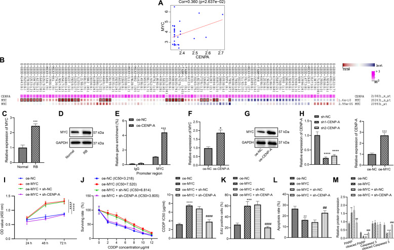
Fig. 3. CENPA promotes the expression of Myc, and reverses the inhibitory effect of Myc on the chemosensitivity of WERI-RB-1/CDDP cells.
A The positive correlation between CENPA and Myc identified through the analysis of microarray GSE28133. B The co-expression between CENPA and Myc identified by MEM analysis. C The differential expression of Myc in retinoblastoma determined by RT-qPCR. D The differential expression of Myc in retinoblastoma determined by western blot analysis (*p < 0.05 vs. normal tissues). E The binding of CENPA to Myc promoter region determined by ChIP assay. F The expression of Myc determined by RT-qPCR. G The expression of Myc determined by western blot analysis. H CENPA silencing and Myc overexpression efficiency determined by RT-qPCR (*p < 0.05 vs. cells transfected with oe-NC or sh-NC). I Cell viability assessed by CCK8 assay. J IC50 value determined by CCK8 assay. K Cell proliferation assessed by EdU assay. L Cell apoptosis assessed by flow cytometry. M Expression of PARP, Cleaved PARP, caspase 3, and Cleaved caspase 3 in cells after transfection determined by western blot analysis. *p < 0.05 vs. cells transfected with oe-NC; #p < 0.05 vs. cells co-transfected with oe-Myc + sh-NC. */#p < 0.05; **/##p < 0.01; ***/###p < 0.001; ****/####p < 0.0001. The experiment was independently repeated three times.

