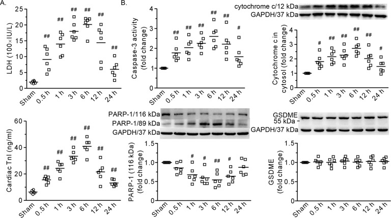
Fig. 1. A time course of the effects of severe burns on the heart.
A Plasma levels of LDH and cTnI. B The results of caspase-3 activity and representative blots and densitometric analysis of cytochrome c, PARP-1, and GSDME in the left ventricle. GAPDH served as a loading control. 0.5–24 h refers to the time point at which the heart specimens were harvested after burn injury. Each independent data with the mean is presented. n = 6 rats in each group. Western blotting was performed in six independent biological experiments, and there were three technical replicates per sample. #P < 0.05, ##P < 0.01 vs. the sham group.

