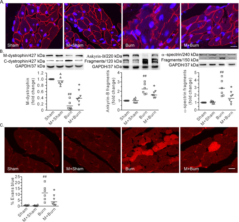
Fig. 5. MDL28170 improved membrane-associated cytoskeleton and plasma membrane integrity in cardiomyocytes after burn.
A Representative confocal images showing dystrophin distribution. Scale bar, 10 μm. B Representative blots showing dystrophin in the membrane and cytosolic fractions, full-length and fragments of ankyrin-B and α-spectrin, and the densitometric analysis results. GAPDH served as a loading control. The ratio of the membrane level to total level or the ratio of the fragment to the total signal per lane is normalized to that in the sham group. C Representative images showing cell uptake of Evans blue and quantification of the positive area. Scale bar, 10 μm. Each independent data with the mean is presented. n = 6 rats in each group. Western blotting was performed in six independent biological experiments, and there were three technical replicates per sample. ##P < 0.01 vs. the sham group. *P < 0.05 vs. the burn group.

