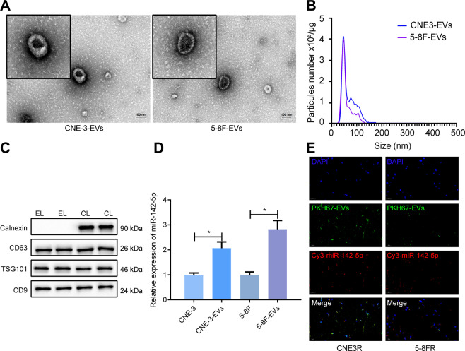
Fig. 3. EVS derived from 5–8 F and CNE-3 cells delivers miR-142-5p into 5-8FR and CNE-3R cells.
A The EVs of NPC cells observed by TEM (scale bar = 100 nm). B The size and concentration of EVs analyzed by NTA. C Western blot to test the EV marker proteins. D The expression of miR-142-5p in 5–8 F and CNE-3 cells and their EVs measured by RT-qPCR. E The localization of PKH67-labeled EVs and Cy3-labeled miR-142-5p in radiotherapy-resistant NPC cells observed by the confocal microscopy. The EVs were collected from parental NPC cell culture medium containing Cy3-labeled miR-142-5p and then used to culture receptor resistant NPC cells for 24 h. *p < 0.05. All experiments were repeated three times.

